

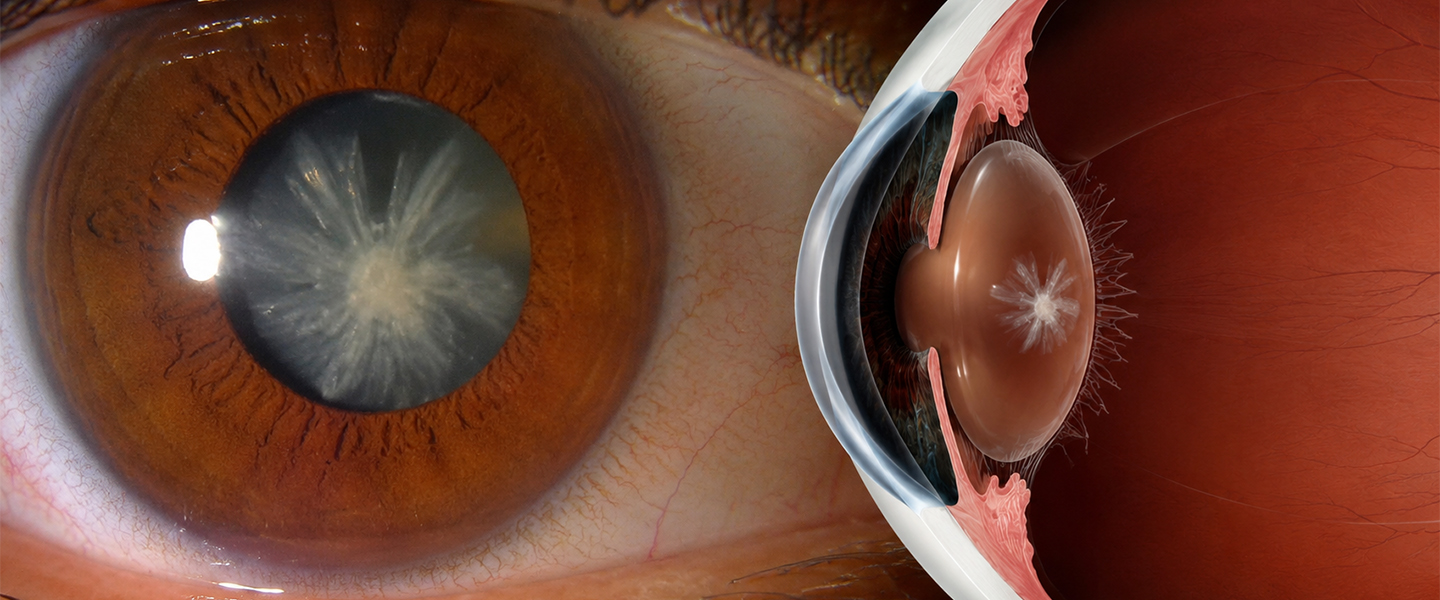
All you need to know about Posterior polar cataract (PPC)
Introduction
Posterior polar cataract (PPC) is a rare form of congenital cataract and is typically inherited as an autosomal dominant condition. Its incidence ranges between 3 to 5 cases per 1000 individuals. PPC is bilateral in 65–80% of cases, and generally, there is no gender predisposition.
Pathogenesis
The position of the lens opacity in PPC is influenced by lens anatomy and the timing of the developmental insult. Suggested causes of PPC include the persistence of the hyaloid artery or invasion of the lens by mesoblastic tissue during embryogenesis. PPC can appear during embryonic life or early infancy, with symptoms often becoming evident between ages 30 to 50. Although the exact pathogenesis is unknown, gene mutations have been linked to PPC, and a positive family history is observed in 40–55% of patients. Key genes associated with PPC include:
- CTTP1 (chromosome 1p36)
- CTTP2 (chromosome 11q22-q22.3)
- CTTP3 (chromosome 20p12-q12)
- CTTP4 (chromosome 10q25)
- CTTP5 (chromosome 14q22-q23)
Clinical Presentation
Signs:
- Abnormal red reflex
- Central posterior discoid plaque on the lens
- Amblyopia (especially in pediatric cases)
Symptoms:
- Asymptomatic in early stages
- Decreased vision
- Photophobia (sensitivity to light)
- Glare
Diagnosis
PPC can be categorized based on clinical presentation:
- PPC with imminent posterior capsule dehiscence
- PPC with preexisting posterior capsule dehiscence
- Spontaneous dislocation
On slit-lamp examination, PPC presents a characteristic “bull’s eye” appearance. Anterior vitreous examination may reveal oil-like droplets, a distinguishing feature in PPC cases.
Classification
Duke and Elders Classification:
- Stationary – Circular, well-defined opacity with a central concentric ring on the posterior capsule.
- Progressive – Characterized by radiating, rider-type opacities in the posterior cortex.
Singh’s Classification
| TYPE 1 | Presence of posterior polar cataract along with posterior subcapsular cataract |
| TYPE 2 | Onion ring-shaped posterior polar cataract with presence or absence of white spot at the edge |
| TYPE 3 | Discrete round or oval opacity at the center with surrounding white spots at the edge. This is usually associated with thin or absent posterior capsule |
| TYPE 4 | Combination of any of the above three along with nuclear sclerosis |
• Schroeder’s Classification further classify PPC in pediatric patients based on the degree of red reflex obstruction through the pupil.
| GRADE 1 | Very small PPC without any effect on the visual acuity through the clear part of the lens |
| GRADE 2 | PPC obstructing two-third part of the pupil |
| GRADE 3 | PPC with the surrounding area of optical distortion. A clear red reflex is appreciated only through a dilated pupil |
| GRADE 4 | Large PPC, no red reflex is observed even through a dilated pupil |
Decision to Operate
Surgery should be deferred as long as possible and is only recommended when the patient experiences difficulty with daily activities. Preoperative counseling is essential, including discussing the risks of nucleus drop, posterior capsular rupture, prolonged operative time, and delayed visual recovery. Nd
capsulotomy may be employed for residual plaque removal. Genetic counseling and family screening are also crucial due to the genetic nature of PPC.
Surgical Approach
Medical management has limited efficacy in PPC. Pediatric patients with PPC should undergo evaluation for systemic syndromes like ectodermal dysplasia, ichthyosis, scleroderma, dyskeratosis congenita, and Rothmund syndrome.
Indications for Surgery:
- Persistent glare and photophobia
- Difficulty reading fine print
- Soft nucleus type PPC or pediatric cases where prompt surgery may prevent blindness
Phacoemulsification is preferred over extracapsular cataract extraction (ECCE) due to lower complication rates. ECCE is generally reserved for cases with a harder cataract and dense central plaque.
Phacoemulsification Procedure:
- Incision – A coaxial corneal or scleral incision is made, or a microincision for a bimanual technique.
- Capsulorhexis – Should not exceed 5–5.5 mm to ensure adequate support for a sulcus-fixated intraocular lens in case of posterior capsule rupture.
- Hydroprocedure – Hydrodissection is avoided, while hydrodelineation is performed in multiple quadrants using minimal fluid to separate the endonucleus from the epinucleus, minimizing disturbance of the posterior capsule.
- Inside-out Hydrodelineation – A trench is sculpted, and fluid is directed perpendicularly through one wall of the trench to separate lens fibers.
- Rotation – Avoid rotating the nucleus to prevent posterior capsule rupture.
- Nucleotomy – Bimanual cracking and division of the nucleus are performed with care to avoid capsular distortion.
- Epinucleus Removal – The central plaque is addressed last to prevent fragments from entering the vitreous cavity.
Posterior Capsular Dehiscence Management: Early recognition and prompt management of posterior capsule rupture are essential for good visual outcomes.
IOL Implantation: Before intraocular lens (IOL) implantation, a viscoelastic substance is injected to expand the capsular bag. The IOL is carefully positioned to avoid contact with the compromised posterior capsule.
Modified Epinucleus Prechop for Dense PPC:
This technique involves segmenting the anterior epinucleus in situ before emulsifying the dense endonucleus and then removing the posterior epinucleus and polar plaque.
Summary
Posterior polar cataract poses unique surgical challenges due to its anatomical complexity. Successful outcomes rely on meticulous technique, including:
- Controlled capsulorhexis
- Avoidance of cortical-cleaving hydrodissection
- Gentle hydrodelineation
- Atraumatic nucleus handling
- Addressing the central epinuclear plate last during cleanup
Patience and precision are critical in achieving favorable postoperative results.
References
1. Birkholz ES, Oetting TA, Kitzmann AS. Posterior Polar Cataract. EyeRounds.org. Available from EyeRounds.
2. Smita Anand, Nimmi Rani. “Posterior Polar Cataract and Its Management,” Surgical Science, Vol.6 No.7, 2015.
3. NCBI – Posterior Polar Cataract
4. Vasavada AR, Vasavada VA. Managing the posterior polar cataract: An update. Indian J Ophthalmol. 2017 Dec;65(12):1350-1358.
5. Kalantan H. Posterior polar cataract: A review, Saudi Journal of Ophthalmology, Volume 26, Issue 1, 2012, Pages 41-49.
6. American Academy of Ophthalmology – Posterior Polar Cataract